Nanoluminofors based on siliсates and germanates of rare earth elements for visualization of biotissues
Abstract
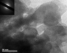
Nanoparticles of silicates and germanates with a general formula Sr2R8–x–yErxYbyM6O26 (R = Y, La; M = Si, Ge) were produced in vacuum by the method of pulse electron beam evaporation. An upconversion photoluminescence of the nanoparticles was detected during the excitation by a laser with a wavelength of 980 nm with a predominance of lines in the red and near infrared regions of the spectrum. Due to their optical properties, the nanoparticles can be excited directly through the biotissues to visualize various pathologies. The obtained nanosamples have K-jumps of X-ray radiation absorption in the 10−100 keV energy region. This opens up prospects for the use of the nanoparticles as X-ray contrast agents. Thus, the nanoparticles have both optical and X-ray contrast characteristics, and therefore have the potential necessary for imaging and diagnosing pathologies in biological tissues.
Keywords
Full Text:
PDFReferences
Erathodiyil N, Ying JY. Functionalization of inorganic nanoparticles for bioimaging applications. Acc Chem Res. 2011;44:925–935. doi:10.1021/ar2000327
Kim J, Piao Y, Hyeon T. Multifunctional nanostructured materials for multimodal imaging, and simultaneous imaging and therapy. Chem Soc Rev. 2009;38:372–390. doi:10.1039/b709883a
Wang G, Peng Q, Li Y. Lanthanide-doped nanocrystals: syn-thesis, optical-magnetic properties, and applications. Acc Chem Res. 2011;44:322–332. doi:10.1021/ar100129p
Zeng S, Yi Z, Lu W, Qian C, Wang H, Rao L, Zeng T, Liu H, Liu H, Fei B, Hao J. Simultaneous realization of phase/size manipulation, up conversion luminescence enhancement, and blood vessel imaging in multifunctional nanoprobes through transi-tion metal Mn2+ doping. Adv Funct Mater. 2014;24:4051–4059. doi:10.1002/adfm.201304270
Liu Y, Meng X, Bu W. Up conversion-based photodynamic cancer therapy. Coord Chem Rev. 2019;379:82–98. doi:10.1016/j.ccr.2017.09.006
Wang J, Wang F, Wang C, Liu Z, Liu X. Single-band up conversion emission in lanthanide-doped KMnF3 nanocrystals. Angew Chem Int Ed Engl. 2011;50(44):10369–10372. doi:10.1002/anie.201104192
Woźny P, Runowski M, Lis S. Emission color tuning and phase transition determination based on high pressure upconversion luminescence in YVO4: Yb3+, Er3+ nanoparticles. J Lumin. 2019;209:321–327. doi:10.1016/j.jlumin.2019.02.008
Sun Y, Zhu X, Peng J, Li F. Core–shell lanthanide up conversion nanophosphors as four-modal probes for tumor angiogenesis imaging. ACS Nano. 2013;7:11290–11300. doi:10.1021/nn405082y
Zhou J, Zhu XJ, Chen M, Sun Y, Li FY. Water-stable NaLuF4-based up conversion nanophosphors with long-term validity for multimodal lymphatic imaging. Biomater. 2012;33:6201–6210. doi:10.1016/j.biomaterials.2012.05.036
Liu Q, Sun Y, Li C, Zhou J, Li C, Yang T, Zhang X, Yi T, Wu D, Li F. 18F-Labeled magnetic-up conversion nanophosphors via rare-Earth cation-assisted ligand assembly. ACS Nano. 2011;5:3146–3157. doi:10.1021/nn200298
Li Z, Zhang Y, Huang L, Yang Y, Zhao Y, El-Banna G, Han G. Nanoscale “fluorescent stone”: luminescent calcium fluoride nanoparticles as theranostic platforms. Theranostics. 2016;6(13): 2380–2393. doi:10.7150/thno.15914
Zuev MG, Larionov LP, Strekalov IM. New radiopaque contrast agents based on re tantalates and their solid solutions. SOP Trans Phys Chem. 2014;1(2):53–64. doi:10.15764/pche.2014.02005
Sokovnin SYu, Il'ves VG, Zuev MG. Production of complex metal oxide nanopouders using pulsed electron beam in low-pressure gas for biomaterials application, «Engineering of Nanobiomaterials Applications of Nanobiomaterials» Elsevier. 2016;2 (Chapter 2). Edited by Alexandru Grumezescu. P. 29–76. doi:10.1016/B978-0-323-41532-3.00002-6
Zuev MG, Larionov LP. biomedical material for diagnosing pathologies in biological tisssues. Patent RU 2734957.
Wang C, Tao H, Cheng L, Liu Z. Near-infrared light induced in vivo photodynamic therapy of cancer based on up conversion nanoparticles. Biomater. 2011;32:6145–6154. doi:10.1002/adfm.20130427
Lv Y, Jin Y, Sun T, Su J, Wang C, Ju G, Chen Li, Hu Y. Visible to NIR down-shifting and NIR to visible upconversion luminescence in Ca14Zn6Ga10O35:Mn4+, Ln3+ (Ln=Nd, Yb, Er). Dyes Pig-ments. 2019;161:137–146. doi:10.1016/j.dyepig.2018.09.052
Huang Z, Xu H, Meyers AD, Iusani A, Wang L, Tagg R, Barqawi AB, Chen YK. Photodynamic therapy for treatment of solid tumors-potential and technical challenges. Technol Cancer Res Treat. 2008;7(4):309–320. doi:10.1177/153303460800700405
Liqiang L, Lanlan D, Weijia Z, Jiawei S,Xiaoqiang Y, Wei L. A phthalocyanine based nearinfrared photosensitizer: Synthesis and in vitro photodynamic activities. Bioorg Med Chem Lett. 2013;23:3775–3779. doi:10.1016/j.bmcl.2013.04.093
Zhou J, Sun Y, Du X, Xiong L, Hua H, Li F. Dual-modality in vivo imaging using rare-earth nanocrystals with nearinfrared to near-infrared (NIR-to-NIR) upconversion luminescence and magnetic resonance properties. Biomater. 2010;31:3287–3295. doi:10.1016/j.biomaterials.2010.01.040
Chantler CT. X-Ray form factor, attenuation and scattering tables. J Phys Chem Ref Data. 2000;29:597–1048. doi:10.18434/T4HS32
DOI: https://doi.org/10.15826/chimtech.2022.9.2.S12
Copyright (c) 2022 Mikhail G. Zuev, Vladislav G. Ilves, Sergei Yu. Sokovnin

This work is licensed under a Creative Commons Attribution 4.0 International License.
Chimica Techno Acta, 2014–2025
eISSN 2411-1414
Copyright Notice


